

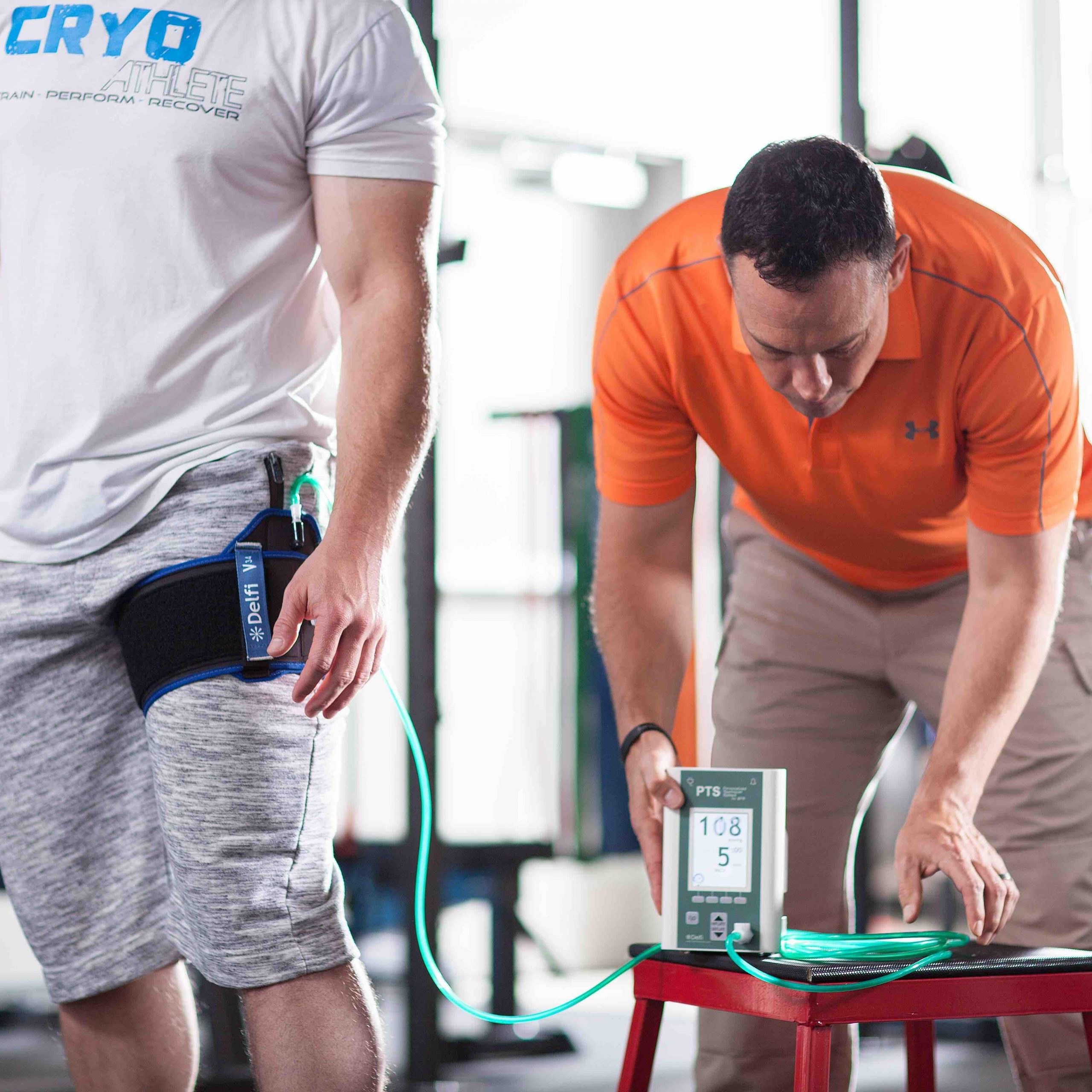
Johnny Owens, PT, first developed blood flow restriction (BFR) training at Brooke Army Medical Center to improve limb strength of wounded warriors without compromising vulnerable soft tissue and joint structures.1 BFR training is typically performed with a tourniquet system that is similar to a blood pressure cuff on the proximal upper and/or lower extremities.2-6 The cuff is typically pressurized from 120-230 mmHg, so as to impede venous blood return while maintaining arterial flow, leading to an accumulation of exercise metabolites and an exaggerated physiologic training response at lower loads.2-6
While the American College of Sports Medicine (ACSM) and the National Strength and Conditioning Association (NSCA) recommend training at 70-80% of one repetition maximum (1RM) to optimize muscle strength and hypertrophy, BFR is typically performed at 20% to 50% of 1RM.6 Similarly, aerobic fitness is traditionally prescribed at 75% of VO2 max, but BFR aerobic training is typically performed at 20% of VO2 max.7, 8 As such, BFR training may provide a significant advantage for maximizing physical therapy outcomes and athletic performance. As Fry et al. points out, “In the last 8 to 10 years, low-load training with blood flow restriction has attracted a lot of attention, both as a possible alternative to heavy resistance exercise in the rehabilitation setting and as a training method to increase muscle strength and size in healthy individuals.“3
SAFETY CONSIDERATIONS
Given that tourniquets control blood flow via general occlusion, the risk of vessel, nerve and skin injury must be considered. Odinsson et al. reviewed 63,484 surgical procedures, whereby tourniquets were required to manage blood flow.9 Despite tourniquets being inflated to over 100 mmHg (on average) above systolic blood pressure, most surgeons safely apply them for 2 hours (on average) with a minimal complication rate of 0.04%.9 As a comparison, BFR training is typically performed with less pressure for a duration of about 10-15 minutes,10 which is approximately one eighth of the surgical occlusion times reported by Odinsson et al.9 To further increase safety during BFR training, a number of investigators recommend wider cuffs than those used during surgery, as there is an inverse relationship between cuff width and occlusive pressure.11, 12
Clark et al. also measured vascular stiffness and abnormal blood clotting via D-Dimer testing during a single bout of BFR exercise and over a 4-week course of BFR training and reported no change.10 Moreover, the authors found no difference in nerve conduction velocity during BFR training.10 Rather, Poton and Polito found lower values of systolic blood pressure, diastolic blood pressure and heart rate following low-intensity, BFR training compared with more traditional, high-intensity strength work.13 In a systematic review of safety issues related to BFR training, Loenneke et al. evaluated cardiovascular stress, oxidative stress, muscle damage and nerve conduction and concluded that BFR is a safe training alternative for individuals regardless of age and training status when used in a controlled environment by trained and experienced personnel.14
STRENGTH TRAINING
Low-intensity, blood flow restriction training is thought to create an environment of low-oxygen availability, causing the activation of type II muscle fibers and anaerobic metabolism.8, 15 The anaerobic environment leads to a build-up of muscle metabolites, which stimulate a number of changes required for muscle and bone strengthening.16 Together, the hypoxic environment and metabolites stimulate neural afferents, causing a significant increase in growth factor hormone (GFH).17 While Takarada reported a 290 times increase in GFH after a single bout of BFR exercise (5 sets of bilateral knee extensions to fatigue, at 20% of 1RM with an average 214 mmHg of cuff pressure) compared to baseline,18 Pierce et al. used a similar protocol and found a lower, but still physiologically relevant, 9-fold increase.8, 19
Fry et al. also reported a 9-fold increase in GFH compared to baseline following a single session of BFR exercise session (one set of 30 repetitions of bilateral leg extensions at 200mmHg cuff pressure, followed by 3 sets of 15 repetitions with 30 second rest intervals between sets).3 While a number of studies have also measured changes in insulin-like growth hormone17 and testosterone20 following blood flow restriction training, the impact of BFR training on these hormones remains controversial.8 Moreover, West et al. did not find a correlation between testosterone, growth hormone and insulin-like growth hormone and muscle protein synthesis following BFR training.21 This finding suggests that local instead of systemic events may predominantly drive physiologic changes post BFR training.8
Local hypoxia and metabolites secondary to BFR training result in a 3-fold increase in ribosomal protein S6 kinase beta-1, a product of the rapamycin (mTOR) pathway, which is responsible for stimulating muscle protein synthesis (MPS), and subsequently, hypertrophy.22 8-hours of BFR training has also shown to decrease proteolytic gene pathways 2-fold compared to traditional high-load exercise, suggesting an increase in muscle remodeling.23 In addition, after an 8 week, low-intensity BFR program, Laurentino et al. reported a down regulation of myostatin mRNA, consistent with traditional, high-intensity strength training programs.24 Given that myostatin is a negative growth factor that inhibits satellite cells and is inversely related to MPS, low-intensity-BFR may be just as effective as traditional, high-load strengthening program for muscle growth.8
It is perhaps worth noting that the effect size of for muscle hypertrophy and strength secondary to low-load, BFR training (as compared to low-load strength training, alone), is .39 and .58, respectively.25 Moreover, it takes an average of 6 weeks of traditional, high-intensity strength training to transition from neural adaptation to muscle hypertrophy.26 However, muscle strength and hypertrophy following low-load, BFR has been shown to occur during the first four weeks of training, suggesting that working smarter instead of harder might truly be a better strategy.8, 26
MUSCLE ATROPHY
Following an injury or surgery, muscle weakness and atrophy are often an issue secondary to disuse and immobilization. After 5 and 14 days of leg immobilization, Wall et al. reported a 3.5% and 8.4% decrease in muscle cross sectional area, a 1.4% and 3.1% reduction in strength, a 68% and 54% decrease in strength and a 68% and 54% increase in myostatin expression, respectively.27 While pain and inflammation make traditional therapeutic exercise challenging, if not impossible, in this patient population, BFR in the absence of muscle contraction may provide an alternative training strategy. Following 2 weeks of lower extremity immobilization, subjects that received BFR (5 repetitions of 5 minutes at 200mmHg with 3 minutes rest between sets) demonstrated less knee and ankle muscle strength loss than non-active subjects and subjects performing isometric exercises.28 Patients that undergo anterior cruciate ligament reconstruction surgery typically experience a significant reduction in quadriceps cross sectional area. However, Takarada et al. reported a 9.4% cross sectional area reduction following BFR (5 repetitions of 5 sets of lower extremity limb occlusion at 238mmHg for 5 minutes with 3 minutes rest between sets) compared to a 20.7% reduction in patients receiving placebo BFR.29 Thus, BFR training has the potential to significantly improve therapeutic outcomes following a traumatic injury and/or surgery.
BONE HEALTH
There is also some evidence that BFR training may be beneficial for bone health. Following 2 days of BFR consistent with Takarada et al. and at a cuff pressure 40% greater than the systolic blood pressure measured in the upper extremity, young-male subjects experienced a significant reduction in the bone reabsorption marker serum N-terminal cross linking telopeptide of type 1 collagen compared to those receiving only low-intensity exercise.30 In addition, the authors reported a 21% increase in serum bone-specific alkaline phosphatase, a marker of bone formation, in older male subjects, compared to low-intensity exercise alone.30 While the mechanism of bone strengthening secondary to BFR is not fully understood, Loenneke et al. suggests that it may be due to “increased interstitial fluid flow-induced sheer stress within the osteocyte membrane”.8, 25, 31
AEROBIC TRAINING
BFR aerobic training is typically performed via walking or cycling with pressure cuffs set to 200 mmHg over the proximal lower extremities, bilaterally.8 After 3-weeks of a BFR walking program at 20-40% of VO2 max, performed twice daily, Abe et al. reported a significant improvement in lower extremity size and strength compared to subjects performing the same program without BFR.32 In a separate study, Abe found that participants completing a 15-minute BFR cycling program at 40% VO2 max, 3 times per week, experienced improved lower extremity muscle size and strength along with a 6.4% improvement in VO2 max.8, 33 Yet, participants that cycled at the same intensity for 40 minutes, 3 times weekly, did not improve on any of the fitness-related variables.33
CONCLUSIONS
Clearly, BFR training may be a valuable tool in the physical therapy profession, as it may facilitate a safer and more efficient strategy for making patients stronger.8 It also may help improve cardiovascular performance and overall bone health.8 Thus, BFR training may lead to enhanced clinical outcomes in patients suffering from acute, subacute and chronic musculoskeletal issues. While not appropriate for every patient, BFR training may also by a particularly powerful tool for patients suffering from disuse, atrophy and/or general deconditioning.34
The American Academy of Manipulative Therapy Fellowship is fortunate to have a strong relationship with champion power lifter and world record holder, Donnie Thompson. During the AAMT Fellowship in Orthopaedic manual Physical Therapy, Mr. Thompson provides training on strength progression, nutrition and a number of alternative manual therapy strategies such as blood flow restriction. As Mr. Thompson often reminds the AAMT fellows-in-training, the best way to treat an injury is to prevent it. In this regard, the use of BFR training to maximize the strength of muscle, bone and connective tissue—so that it can withstand more force and be less susceptible to injury—should be considered part of injury prevention and/or rehabilitation programs.
AUTHORS
Dr. Cameron Moore, DPT, Cert. SMT, Cert. DN, FAAOMPT, Dip. Osteopractic
Fellow, AAMT Fellowship in Orthopaedic Manual Physical Therapy
Dr. Joe Ciccone, DPT, SCS, Cert. SMT, Cert. DN, FAAOMPT, Dip. Osteopractic
Fellow, AAMT Fellowship in Orthopaedic Manual Physical Therapy
Columbia University Medical Center, New York, NY
Dr. Raymond Butts, PhD, DPT, MSc, Cert. SMT, Cert. DN, Dip. Osteopractic
Senior Faculty, American Academy of Manipulative Therapy Fellowship, Atlanta, GA
REFERENCES
- Blood Flow Restriction and Physical Therapy. (2015, January). Move Forward PT: Physical Therapists Bring Motion to Life. Retrieved July 17, 2017, from https://www.moveforwardpt.com/Radio/Detail/blood-flow-restriction-training-physical-therapy
- Wernbom M, Jarrebring R, Andreasson MA, Augustsson J. Acute effects of blood flow restriction on muscle activity and endurance during fatiguing dynamic knee extensions at low load. J Strength Cond Res. 2009;23(8):2389-95.
- Fry CS, Glynn EL, Drummond MJ, Timmerman KL, Fujita S, Abe T, et al. Blood flow restriction exercise stimulates mTORC1 signaling and muscle protein synthesis in older men. J Appl Physiol (1985). 2010;108(5):1199-209.
- Loenneke JP, Thiebaud RS, Fahs CA, Rossow LM, Abe T, Bemben MG. Blood flow restriction: effects of cuff type on fatigue and perceptual responses to resistance exercise. Acta Physiol Hung. 2014;101(2):158-66.
- Kim D, Singh H, Loenneke JP, Thiebaud RS, Fahs CA, Rossow LM, et al. Comparative Effects of Vigorous-Intensity and Low-Intensity Blood Flow Restricted Cycle Training and Detraining on Muscle Mass, Strength, and Aerobic Capacity. J Strength Cond Res. 2016;30(5):1453-61.
- Vilaça-Alves J, Neto G, Morgado N. Acute Effect of Resistance Exercises Performed by the Upper and Lower Limbs with Blood Flow Restriction on Hemodynamic Responses. J Exerc Physiol Online. 2016;19(3):100-9.
- Seiler S. What is best practice for training intensity and duration distribution in endurance athletes? Int J Sports Physiol Perform. 2010;5(3):276-91.
- Hackney KJ, Everett M, Scott JM, Ploutz-Snyder L. Blood flow-restricted exercise in space. Extrem Physiol Med. 2012;1(1):12.
- Odinsson A, Finsen V. Tourniquet use and its complications in Norway. J Bone Joint Surg Br. 2006;88(8):1090-2.
- Clark BC, Manini TM, Hoffman RL, Williams PS, Guiler MK, Knutson MJ, et al. Relative safety of 4 weeks of blood flow-restricted resistance exercise in young, healthy adults. Scand J Med Sci Sports. 2011;21(5):653-62.
- Crenshaw AG, Hargens AR, Gershuni DH, Rydevik B. Wide tourniquet cuffs more effective at lower inflation pressures. Acta Orthop Scand. 1988;59(4):447-51.
- Graham B, Breault MJ, McEwen JA, McGraw RW. Occlusion of arterial flow in the extremities at subsystolic pressures through the use of wide tourniquet cuffs. Clin Orthop Relat Res. 1993(286):257-61.
- Poton R, Polito MD. Hemodynamic response to resistance exercise with and without blood flow restriction in healthy subjects. Clin Physiol Funct Imaging. 2016;36(3):231-6.
- Loenneke JP, Wilson JM, Wilson GJ, Pujol TJ, Bemben MG. Potential safety issues with blood flow restriction training. Scand J Med Sci Sports. 2011;21(4):510-8.
- Krustrup P, Soderlund K, Relu MU, Ferguson RA, Bangsbo J. Heterogeneous recruitment of quadriceps muscle portions and fibre types during moderate intensity knee-extensor exercise: effect of thigh occlusion. Scand J Med Sci Sports. 2009;19(4):576-84.
- Suga T, Okita K, Morita N, Yokota T, Hirabayashi K, Horiuchi M, et al. Intramuscular metabolism during low-intensity resistance exercise with blood flow restriction. J Appl Physiol (1985). 2009;106(4):1119-24.
- Takano H, Morita T, Iida H, Asada K, Kato M, Uno K, et al. Hemodynamic and hormonal responses to a short-term low-intensity resistance exercise with the reduction of muscle blood flow. Eur J Appl Physiol. 2005;95(1):65-73.
- Takarada Y, Nakamura Y, Aruga S, Onda T, Miyazaki S, Ishii N. Rapid increase in plasma growth hormone after low-intensity resistance exercise with vascular occlusion. J Appl Physiol (1985). 2000;88(1):61-5.
- Pierce JR, Clark BC, Ploutz-Snyder LL, Kanaley JA. Growth hormone and muscle function responses to skeletal muscle ischemia. J Appl Physiol (1985). 2006;101(6):1588-95.
- Loenneke JP, Wilson JM, Pujol TJ, Bemben MG. Acute and chronic testosterone response to blood flow restricted exercise. Horm Metab Res. 2011;43(10):669-73.
- West DW, Kujbida GW, Moore DR, Atherton P, Burd NA, Padzik JP, et al. Resistance exercise-induced increases in putative anabolic hormones do not enhance muscle protein synthesis or intracellular signalling in young men. J Physiol. 2009;587(Pt 21):5239-47.
- Fujita S, Abe T, Drummond MJ, Cadenas JG, Dreyer HC, Sato Y, et al. Blood flow restriction during low-intensity resistance exercise increases S6K1 phosphorylation and muscle protein synthesis. J Appl Physiol (1985). 2007;103(3):903-10.
- Manini TM, Vincent KR, Leeuwenburgh CL, Lees HA, Kavazis AN, Borst SE, et al. Myogenic and proteolytic mRNA expression following blood flow restricted exercise. Acta Physiol (Oxf). 2011;201(2):255-63.
- Laurentino GC, Ugrinowitsch C, Roschel H, Aoki MS, Soares AG, Neves M, Jr., et al. Strength training with blood flow restriction diminishes myostatin gene expression. Med Sci Sports Exerc. 2012;44(3):406-12.
- Loenneke JP, Wilson JM, Marin PJ, Zourdos MC, Bemben MG. Low intensity blood flow restriction training: a meta-analysis. Eur J Appl Physiol. 2012;112(5):1849-59.
- Moritani T, deVries HA. Neural factors versus hypertrophy in the time course of muscle strength gain. Am J Phys Med. 1979;58(3):115-30.
- Wall BT, Dirks ML, Snijders T, Senden JM, Dolmans J, van Loon LJ. Substantial skeletal muscle loss occurs during only 5 days of disuse. Acta Physiol (Oxf). 2014;210(3):600-11.
- Kubota A, Sakuraba K, Sawaki K, Sumide T, Tamura Y. Prevention of disuse muscular weakness by restriction of blood flow. Med Sci Sports Exerc. 2008;40(3):529-34.
- Takarada Y, Takazawa H, Ishii N. Applications of vascular occlusion diminish disuse atrophy of knee extensor muscles. Med Sci Sports Exerc. 2000;32(12):2035-9.
- Takarada Y, Sato Y, Ishii N. Effects of resistance exercise combined with vascular occlusion on muscle function in athletes. Eur J Appl Physiol. 2002;86(4):308-14.
- Fritton SP, Weinbaum S. Fluid and Solute Transport in Bone: Flow-Induced Mechanotransduction. Annu Rev Fluid Mech. 2009;41:347-74.
- Abe T, Kearns C, Fujita S, Sakamaki M, Sato Y, Brechue W. Skeletal muscle size and strength are increased following walk training with restricted blood flow: implications for training duration and frequency. Int J KAATSU Train Res. 2009;5:9-15.
- Abe T, Fujita S, Nakajima T, Sakamaki M, Ozaki H, Ogasawara R, et al. Effects of Low-Intensity Cycle Training with Restricted Leg Blood Flow on Thigh Muscle Volume and VO2MAX in Young Men. J Sports Sci Med. 2010;9(3):452-8.
- Heitkamp HC. Training with blood flow restriction. Mechanisms, gain in strength and safety. J Sports Med Phys Fitness. 2015;55(5):446-56.
